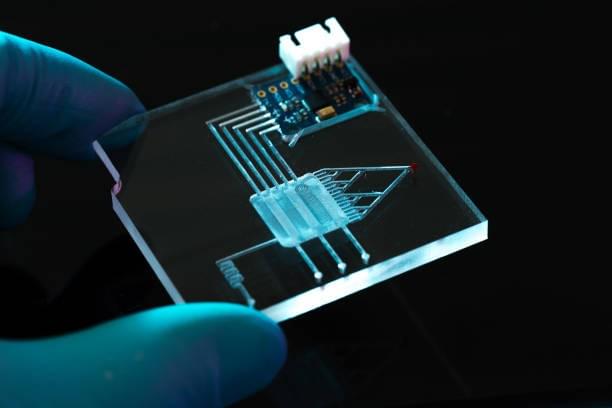
The microfluidic chamber is an excellent tool to study the behavior of cells in aqueous environments. Unlike conventional plates, the x-z plane provides a clear view of the entire volume of the sample. The cell nucleus and two beads are circular and in direct contrast to the conventional x-y plane, which shows the same volume during shear flow in the x direction. In addition, the n- and p-planes show the same amount of fluid in the same location. These two images also illustrate the displacement of the nucleus and the beads.
The microfluidic chamber was designed to allow researchers to isolate distinct segments of axons. These devices have revolutionized the neuroscience field. Using the microfluidic platform, researchers can now study axon regeneration and transport, biochemical alterations in axons, and identify the factors that affect them. Because the chamber allows for such detailed analyses, these instruments have become standard tools in hundreds of labs around the world.
The microfluidic chamber uses replica molding to create a multicompartment culture system. The design of this neuron device is easy to produce, even in biology labs without clean rooms. Its high-quality components make it an ideal choice for neuroscience research. This versatile equipment can be used for a variety of purposes, from cell migration to chemotaxis. Its unique properties also make it possible to perform studies on the motion of neutrophils and other cell types.
The mLP chamber has the advantage of allowing researchers to investigate calcium dynamics both locally and globally. In the axonal compartment, glutamate perfusion led to a slower increase in calcium than vehicle perfusion, whereas the vehicle-perfused dendrite showed a rapid increase in calcium. Furthermore, the gray layers are suitable for growing axons, which can be photographed with a microscope.
The microfluidic chamber is a useful tool for high-throughput drug screening and 3D cell culture. The inlet of the cell chamber must be blocked whenever the drug is changed. The inlet must be blocked when switching the drugs. In the x-y plane, the confocal image of the bead-coated glass is used to calculate the height of the beads in the cell chamber. The z-y plane, on the other hand, can be used to determine the distance between the bead and the glass.
This chamber has the potential for multiple uses. For example, it can be used for imaging neuron/glia co-culture. In addition, it can be used for experiments involving different drugs. Moreover, it can be utilized for determining the shear modulus of various tissues. Its complexities make it a highly versatile tool for research. The device has many advantages. Besides, it allows scientists to observe neuronal cells and internal organelles simultaneously.
This link https://en.wikipedia.org/wiki/Paper-based_microfluidics sheds light into the topic—so check it out!